
Talus Bio's Ptarmigan-1, the Human Cancer Models Initiative, and Vilya Research's Vilya-2
Kiin Bio's Weekly Insights

Welcome back to your weekly dose of AI news for Life Science! This weeks fix:
Talus Bio threw out 3D structure entirely and screened 3.4 billion compounds against the full human proteome in under a day, and it works on targets that co-folding models cannot even attempt.
Nature published three coordinated papers dropping over 1,200 new cancer models into public repositories, and the buried finding that growth medium alone reshapes which genes look essential should send a shiver down the spine of anyone who trusts a single CRISPR screen.
Vilya Research extended their macrocycle structure model to peptide-protein and small-molecule interfaces, and the accuracy numbers on peptide docking put co-folding methods in an uncomfortable position.
You read this newsletter because staying current in AI x life science matters to you, it matters to us too. That's why we're building a free personalised resource for the community: from papers, patents, and clinical trials to c-suite career moves and job opportunities. So you can stay up to date with what actually matters to you.
We want to know: where else are you looking? What's missing? Two minutes of your time will shape what gets built.
Ptarmigan-1: Structure-free virtual screening across the entire human proteome
What It Is
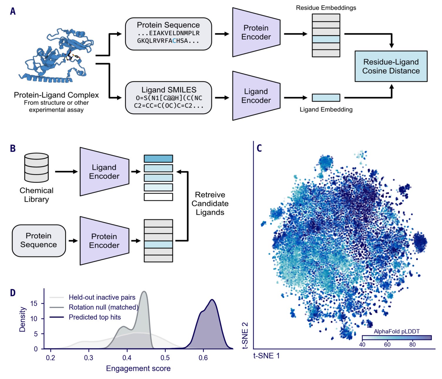
Every virtual screening method in serious use today needs a protein structure. Molecular docking needs a pocket. AlphaFold3, Boltz-2, and Chai-1 need the full 3D complex. For the 87% of human proteins without an approved drug or potent ligand, many of which are disordered or lack a well-defined pocket, that requirement is the bottleneck. Ptarmigan-1 from William Fondrie and Alexander Federation at Talus Bioscience sidesteps it entirely: a contrastive model that co-embeds protein residues and small molecules in a shared latent space from sequence and SMILES alone. No pose, no structure, no pocket definition.
The architecture couples ESM Cambrian for protein sequences with ChemBERTa for compounds, both adapted via LoRA, and trains on chemoproteomic data (activity-based protein profiling, photoaffinity labelling) alongside standard bioactivity databases. Because the data is residue-level, not pocket-level, the model learns where a compound binds, not just whether it binds.
Where This Fits
The speed difference is staggering. Ptarmigan-1 scores a compound in 10 milliseconds. Boltz-2 takes 54 seconds for the same pair. That 5,000-fold gap meant Talus could screen 3.4 billion compounds from the OnePot CORE library against all 20,431 human proteins in under a day on 20 H100 GPU-hours. Try that with co-folding and you are looking at thousands of GPU-years.
On the standard LIT-PCBA benchmark of well-folded, orthosteric targets, Ptarmigan-1 ranked second behind Boltz-2 and ahead of docking tools like Glide-SP and Gnina. Respectable for a method that never sees 3D coordinates. Where it separates from the field is on targets those methods cannot handle: intrinsically disordered proteins where AlphaFold pLDDT drops below 50, cryptic pockets that only open on ligand binding, and covalent sites. On disordered targets, Boltz-2 fell to near-random while Ptarmigan-1 retained positive enrichment across the board.
Why This Is Cool
The STAT6 inhibitor recovery is the part that convinced me this is real. Talus withheld a patent-only inhibitor series from training, screened for it blind, and Ptarmigan-1 recovered the actives and correctly localised them to the engagement residues. A co-folding baseline mislocalised them. That is a practical drug-discovery validation, not a benchmark game.
I think the broader implication is simple: if your target has no structure, or its structure misleads pose-based methods, this is currently the only game in town at proteome scale. It will not replace co-folding for well-characterised targets where pose accuracy matters. It does not give you a binding mode. What it gives you is a way to ask “does anything in this 3-billion-compound library hit my difficult target?” and get an answer in hours rather than never.
Code is not yet publicly available.
Paper: Fondrie et al., bioRxiv 2026
The Human Cancer Models Initiative: 1,200 new models across three coordinated Nature papers
Why This Is Cool
I want to start with the finding that will bother people most: growth medium independently shapes gene essentiality profiles. Not genetic background, not tissue of origin. The medium. The Broad team showed that 3D organoid models revealed WNT pathway dependencies that were completely silenced when the same cells grew in standard 2D culture. If you have been running CRISPR screens exclusively in adherent lines and treating the results as ground truth, these papers suggest you have been measuring an artefact of your culture conditions as much as the biology of your cancer.
What It Is
Nature published three papers simultaneously from the NCI Human Cancer Models Initiative, the Wellcome Sanger Institute, and the Broad Institute’s DepMap programme. Combined, they release over 1,200 new cancer models into public repositories. The specifics:
HCMI contributed 665 models spanning 25 cancer types with 97.8% DNA concordance to their parent tumours, including rare cancers and underrepresented populations historically absent from model collections.
The Sanger team generated 256 colorectal and gastric organoids, paired them with genome-wide CRISPR screens, and identified KRAS allele-specific drug sensitivities that distinguish G12D from G12V mutations at a level standard cell line panels have never captured.
The Broad added 314 NextGen models to DepMap, specifically built to fill gaps in tissue and molecular diversity.
Where This Fits
Cancer model collections are not new. We have had cell line panels from CCLE and organoid banks from the Hubrecht Institute for years. What is new is the scale, the coordination, and the systematic comparison across culture formats. Previous resources let you screen in one system and assume the results translate. These papers explicitly tested that assumption and found it wanting. The Sanger KRAS result alone, where G12D and G12V alleles show different drug sensitivity profiles, has direct implications for how we stratify patients for targeted therapies.
For computational groups, over a thousand new models with matched genomic data, drug response measurements, and CRISPR screen profiles just landed in public databases. If you are training response-prediction models on the same 50 NCI-60 lines everyone else uses, the excuse for not updating your training set just disappeared.
Papers: HCMI (Nature 2026) | Sanger (Nature 2026) | Broad/DepMap (Nature 2026)
Vilya-2: Accurate structural modelling of chemically diverse molecular interfaces
Where This Fits
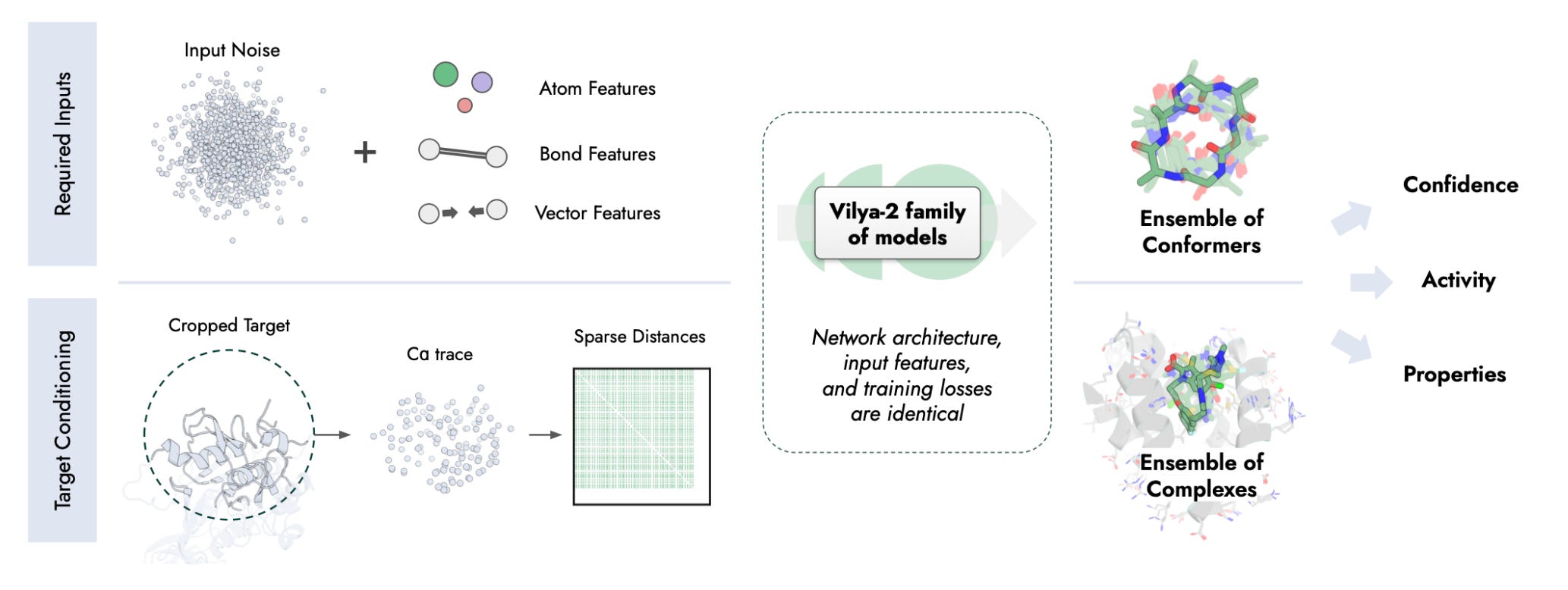
We covered Vilya-1 a couple weeks ago when it launched for macrocycle structure prediction. The model worked well in that narrow lane, but macrocycles are a small corner of molecular design. Vilya-2 is a much more ambitious extension: a diffusion transformer that now handles peptide-protein interfaces, small-molecule docking, and generalisation to molecular classes it was never trained on.
The competitive landscape here is crowded. AlphaFold3, Boltz-2, Chai-1 all do molecular complex prediction. What separates Vilya-2 is an all-atom representation that treats every molecular type the same way, and the resulting accuracy on peptides specifically. 59.1% of peptide interfaces predicted to sub-2 angstrom backbone RMSD is a substantial jump over co-folding methods on the same test set.
What It Is
Pascal Sturmfels and colleagues at Vilya Research built a single model that predicts how molecules sit against protein surfaces. The diffusion transformer generates atomic coordinates for the interface, trained on structural data spanning peptides, small molecules, and macrocycles. Rather than treating each molecular class as a separate problem (which is how most groups have approached this), the unified representation means the model transfers knowledge between classes. It was trained on peptide-protein and small-molecule-protein data and then tested on macrocycles and stapled miniproteins it never saw during training. It got those right too.
On small-molecule docking benchmarks, Vilya-2 reaches state-of-the-art accuracy. On peptide interfaces specifically, it outperforms every co-folding model tested.
Why This Is Cool
Peptide drug design is where I think this matters most immediately. Peptide therapeutics are growing fast, semaglutide being the obvious example, but predicting how a peptide will sit against its target protein has been genuinely hard. Co-folding models were not designed for the extended, flexible interfaces peptides form. Vilya-2 was.
The generalisation result is what makes me think this is more than incremental. A model trained on standard peptides and small molecules that then correctly predicts stapled miniprotein interfaces suggests the representation has learned something transferable about molecular contacts, not just memorised the training set.
Worth watching whether this translates to prospective peptide design. The accuracy is there. The question is whether it holds on targets outside the structural databases it learned from.
Paper: Sturmfels et al., 2025
🗓️ Events & Competitions
The best competitions, hackathons, and community challenges in AI x life sciences, curated weekly. Know something worth featuring? Reply and let us know.
More upcoming events:
BioHackathon Europe 2026 | November 9-13, Barcelona
ELIXIR’s annual international bioinformatics hackathon, running since 2018. 160+ participants, five days of collaborative coding on open bioinformatics infrastructure and tools. The call for project proposals has now closed.
Thanks for reading!
💬 Get involved
We’re always looking to grow our community. If you’d like to get involved, contribute ideas or share something you’re building, fill out this form or reach out to me directly.
Connect With Us
Have questions or suggestions? We'd love to hear from you!
📧 Email Us | 📲 Follow on LinkedIn | 🌐 Visit Our Website